


Equine lameness is a commonly observed condition whose effects are typically concentrated in certain areas of a horse’s anatomy. Understanding in which parts lameness frequently occurs, and properly diagnosing the issues can prove to be a challenging task. This guide aims to shed light on this prevalent issue maintaining a balance between simplicity for everyday understanding and the expertise needed for in-depth comprehension.

Predominant Areas of Lameness
Lameness in horses predominantly surfaces in areas below the knee or hock, with the foot and back tendons being especially susceptible. To comprehend the reason for this frequency, it is essential to understand the anatomy and functionality of these parts.
As a horse’s knee is a complex joint engineered with numerous tiny bones, it effectively distributes and minimises concussion or damaging forces often linked to lameness, hence, having fewer incidents of being affected. The shoulder, another sturdy part of the horse’s anatomy, also experiences lameness infrequently. When it does occur, the impairment usually stems from muscle injury due to sprains or impact blows.
Importantly, below the knee and hock, the horse’s anatomy lacks muscle presence. In this vicinity, the tendons are primarily responsible for bearing strain. If these tendons fail to adequately manage this strain due to weakness or injury, the bone faces increased risk of fracturing, which can result in lameness as well.
Diagnosis of Lameness
The initial step in diagnosing lameness involves pinpointing the pain’s location, which isn’t always apparent. Common practice, when the exact site of lameness remains elusive, is to suspect the foot’s involvement. This advice is rooted in the popular saying, “remove the shoe, even if the horse is lame in the head”. This adage brings to light the foot’s vulnerability owing to the high incidence of troubles that begin there.
However, diagnosing issues related to the foot can be complicated, given the hoof’s tough, horny box conceals the cause of discomfort. It’s often a process of discretion, but through careful examination and understanding of the horse’s anatomy, one can get closer to identifying and resolving the root cause of lameness.
Identifying the Affected Leg
Which leg is affected by lameness? This question has caused quite a bit of confusion and disagreement, and unfortunately, many sound legs have been treated by mistake due to misidentification. The following guidelines will help in understanding and solving this issue.
Initial Indications in the Stable
As mentioned in “Attitude”, a horse may “point” or rest a lame foreleg in front of the other while standing in the stable. Similarly, resting a hind leg is normal. Constant uneasiness on one foot should raise suspicion of lameness.
If the horse doesn’t display any typical signs of lameness while in the stable, the next step is to examine the horse outside on a smooth, hard surface.
Trotting up the Horse
Take the horse out on to a flat, firm surface and make it “trot up,” slowly moving away from and then back toward the examiner. Observe any potential “favoring” of one limb or “dropping” or bearing more heavily on the sound leg. Use the mnemonic “SINKS ON THE SOUND SIDE” to remember this pattern. It’s best to perform this examination immediately after the horse is taken out of the stable, especially after a rest, when stiffness is more pronounced.
Observing Head Nodding
At the trot, head nodding is a reliable indicator of lameness. If the horse is lame in the front, its head will dip as the sound leg touches the ground and rise on the lame one. Severe lameness in both forelegs might give an impression of soundness; however, careful inspection will reveal “pottering” or shortened steps. Additionally, while determining the affected leg, one can often identify the trouble’s location. Turning the leg outwards indicates a problem with the knee or elbow, whereas dragging the toe due to difficulty in forward movement suggests shoulder issues.
Detecting Lameness in Hind Limbs
Lameness in a horse’s hind limb is noticeable through uneven movement of the hindquarters when the horse trots away. The hindquarter on the corresponding side will rise when weight is put on the lame leg, while the opposite quarter will sink down when the sound leg hits the ground (again, remember “SINKS ON THE SOUND SIDE”). Nonetheless, confusion may arise between lameness in a hind limb and the diagonally opposite fore limb if uneven hindquarter movement is not accurately observed.
During the “trot up,” it’s helpful to examine the horse from the side, observing the arc of flight of the feet, stride length, and foot placement on the ground. Often, you may notice that one foot isn’t lifted as high or takes a shorter stride compared to the opposite leg. Be vigilant for any foot landing on the toe or landing more heavily on the heel than normal, as this could denote pain from bearing weight on that area.
Finding the Source of Lameness
Once the affected leg has been identified, it is essential to locate the precise source of the lameness. Several methods can be employed to assist in this process.
Examination by Touch
The affected leg should be felt for heat, tenderness, or enlargements and carefully compared to the sound one. If the leg appears normal, the next step is to remove the shoe and inspect the foot for nails, bruises, corns, and similar issues. Heat in the foot or “pointing” typically indicates that the problem lies in the foot, as does constantly lifting the foot, which suggests significant pain.
Veterinary Examination
A veterinarian will generally examine the limbs in a similar fashion but will also check the joints by carefully flexing and extending each one in turn. They may even hold a joint flexed for a period before trotting the horse up again. This method can make certain joint problems, such as spavin, more noticeable. The vet may also use hoof testers (special pincers) to apply pressure to various parts of the hoof to locate any painful spots.
If the exact location of lameness remains uncertain, a nerve block may be performed. By injecting a local anesthetic around the nerves at a certain level on the limb, the leg is temporarily desensitized below that point. If the seat of lameness is below the injection site, the pain will cease, and the horse will trot up soundly. A nerve block at the pastern will cause a horse with a foot problem to trot soundly. If the horse is still lame, the issue likely originates higher up the leg, and further injections higher up the limb may be necessary to pinpoint the problem.
X-Rays and Blood Tests
If bone damage or a problem within the foot is suspected, it may be necessary to take X-rays to assess the position and extent of the injury. Damaged muscle tissue causes changes in the blood that can be easily detected through blood tests. These tests can be helpful for confirming or ruling out muscle injury as a possible cause of lameness, especially when dealing with back problems.
Causes of Lameness in Horses
Understanding the common causes of lameness in horses can be crucial in addressing the issue effectively and promptly. There are several reasons why a horse might start limping, with the most frequent culprits being strain and damage to the tendons and ligaments, or problems resulting from continuous work on hard surfaces.
Strains and Overexertion
Horses engaged in high-speed activities on soft ground often suffer from strains of the tendons and ligaments. These strains can be exacerbated by poor quality riding, overworking when the horse is already tired, or introducing the horse to an unsuitable “going” (the term that horse people use to describe the condition of the ground or surface on which a horse runs).
In this situation, paying attention to a horse’s fatigue levels and monitoring the terrain used for intense work can be decisive in preventing tendon and ligament strain, ultimately avoiding episodes of lameness.
Long-term work on Hard Surfaces
Working on hard surfaces for extended periods, especially in the case of draught horses, can lead to lameness due to the development of bony enlargements. These enlargements, while they form, are a common cause of discomfort and limping.
In some cases, these bony enlargements appear in and around the joints, like the situation in articular ringbone and spavin, restricting the joints’ movements. Such conditions can result in a stilted action, contributing to the overall lameness.
Check Ligament Injuries
An additional risk from continuous work on hard surfaces is the injury of the “check ligament.” Located with the back tendons between the knee and fetlock, the check ligament enables a horse to sleep while standing. Sprain in this ligament manifests as a painful swelling just below the knee, signifying another potential root of lameness.
By understanding these common causes of lameness, we can further enhance our strategies for horse care, ensuring that our equine companions stay healthy and energetic.
Dislocations
Dislocations, while extremely rare, can cause significant discomfort and compromised mobility in horses. They usually entail ligament and muscle sprains along with the shifting of joints from their original position.
Partial and Complete Dislocations
Partial dislocations are sometimes observed in young horses, specifically at the fetlock or the patella (the “knee-cap” in the horse’s stifle joint). Interestingly, it’s not uncommon for these joints to click back into place on their own accord.
On the contrast, complete dislocation is a serious situation. However, where muscles are more involved than ligaments in holding the joint together, such as in the shoulder joint, the hope for full recovery is more promising.
Symptoms and Treatment of Dislocations
The most common symptoms of a dislocation include observable deformity, loss of use of the limb accompanied by a shortening of it, and often, painful swelling. A trained eye may recognize these symptoms, making it possible to promptly respond to such injuries.
Dislocated bones can occasionally be realigned to their normal position through manipulation under general anesthesia. However, suspected dislocations always warrant immediate veterinary attention. Some cases may require more intensive treatments, and considering the delicacy and complexity of the horse’s physiology, professional help is essential in such situations.
Fractures
Fractures, which can be quite serious injuries in horses, come in various forms, each presenting different challenges and requiring specific treatments for successful healing.
Types of Fractures
As per their nature and form, fractures can be categorized into: simple, compound, comminuted, impacted, and greenstick fractures.
Simple Fractures
Simple fractures present without any external wounds and involve a singular break in the bone. This break can occur across the bone (transverse), at an angle (oblique), or run the full length of the bone (longitudinal).
Compound Fractures
A step further from simple fractures, compound fractures are characterized by an external wound corresponding to the broken bone, making the injury more complex to manage due to the risk of infection.
Comminuted Fractures
Comminuted fractures indicate a bone is shattered into more than two pieces. Reminiscent of a crushing effect, these fractures can be simple, with no external wound, or compound, associated with an open wound.
Impacted Fractures
Impacted fractures occur when the demolished bone sections are driven into each other, creating a compact arrangement that, while painful, does offer some structural stability.
Greenstick Fractures
Greenstick fractures are seen primarily in young horses with more flexible bones. Just like a green stick, which only partially breaks when bent, these fractures involve an incomplete break in the bone.
Symptoms and Detection of Fractures
The primary indicators of fractures typically include pain, loss of function in the affected limb, and subsequent swelling. Unlike dislocations where the joints are ‘fixed’, fractures show movement at the break point. The grating sound and sensation (‘crepitation’) of the broken bone fragments against each other is often evident – a key clue that a fracture may have occurred.
Regardless of the type of fracture, it’s crucial to seek veterinary attention promptly, as these injuries can have serious implications if left untreated, and each fracture type requires specific intervention strategies.
Treatment of Fractures in Horses
The correct treatment of fractures in horses must be based on an understanding of the various fracture types, the specific animal’s condition, and the importance of professional veterinary assistance at the appropriate time.
Immediate Response to Suspected Fractures
When a fracture is suspected, following a fall or accident, a veterinarian should be contacted immediately to examine the horse on the spot before attempting to move the animal. In many instances, attempting to move a horse with an unsupported damaged limb can exacerbate a simple fracture, turning it into a compound or comminuted fracture.
If the injured horse is lying on the ground and struggling to get up, it should be prevented from further exertion. Someone may need to sit or lie across the top of the horse’s neck to hold it down. Placing a coat under its head can safeguard the lower eye from injury, while a second coat over its eyes might help to calm the animal until assistance arrives.
When there’s ample help and equipment available, it may be necessary to use ropes to carefully roll the horse over to facilitate its movement. Thick layers of cotton wool wrapped around the leg with bandages, reinforced by suitable materials (e.g., broom handles or plastic guttering) can provide additional support to the fractured limb.
Both splinting the leg and moving the horse must be carried out under the guidance of a veterinarian. Pain relief and sedation may be required to safely move the horse.
Modern Fracture Treatment Methods
In the past, horses with broken legs were often euthanized to prevent further suffering, as there was no effective method of immobilizing the leg to allow healing. However, advancements in treatment techniques and materials have made it possible to treat certain kinds of fractures in horses more effectively.
Orthopedic plates and screws, combined with fibreglass casts, now render the treatment of some limb fractures similar to that of humans. This has significantly improved the chances of recovery and the quality of life for affected horses.
Treatment Outcomes Based on Fracture Type
- Greenstick fractures usually heal well with rest and immobilization.
- Simple fractures located in the lower parts of the limbs can be successfully treated using surgery and/or fibreglass cast application.
- Comminuted fractures and fractures higher up the leg carry much graver consequences, potentially necessitating euthanasia if there’s no chance of healing.
The most suitable course of action can only be determined after thoroughly assessing the break’s extent and position, typically through an X-ray examination.
Small and Overlooked Fractures
Smaller fractures are often overlooked or misdiagnosed. For example, when X-rays are taken of a swollen joint as part of a lameness examination, small bone fragments—or “chips”—within the joint might be discovered. Similarly, fractures of the splint bones can be mistaken for regular splints, when in fact they are the result of a kick or knock and require surgical intervention. Cracked ribs can also be the product of an injury, such as a kick from another horse, and must be identified and addressed appropriately.
Arthritis in Horses
Arthritis, or Degenerative Joint Disease, is a common issue in horses, particularly as they age, due to the continual wear and tear on their joints from daily activities. Various factors can contribute to the development of arthritis in horses, such as poor conformation, improper shoeing, and frequent exposure to hard surfaces.
Commonly Affected Joints
Arthritic changes are regularly seen in the foreleg fetlock joints, which can become swollen and lead to the formation of articular “windgalls”. The pastern and coffin joints may also be affected, with arthritis in these areas referred to as articular ringbone. In the hock, arthritis can result in spavin.
Symptoms and Treatment of Arthritis
Horses with mild arthritis are typically stiff at first but can warm up during movement. In more severe cases, the horse may become lame. Pain-killing anti-inflammatory drugs have often been used to treat these animals, allowing them to remain sound.
However, in some instances, drugs such as butazolidin (“bute”) can mask the pain so effectively that horses who should be resting continue to be worked, causing further joint damage. In other cases, older and stiffer horses may benefit from such medication.
Innovative Treatments for Degenerative Joint Disease
Newer drugs have been developed to address degenerative joint diseases in horses. These treatments can be injected directly into the affected joint, promoting healing of arthritic changes. By focusing treatment on the specific joint rather than the entire horse, the risk of side-effects that can occur when administering anti-inflammatory drugs orally is reduced.
Management of Arthritic Horses
For horses prone to arthritis, engaging in regular exercise can often help alleviate their symptoms. This typically involves small amounts of work daily rather than less frequent, more intense sessions.
Arthritis is highly prevalent in older horses and tends to worsen during cold and wet weather. As a result, it may be necessary to provide warm shelter or stable older horses during winter months to offer relief from their arthritic pain.
Dealing with Sprains in Horses
Sprains are a common issue among horses. They occur when the ligaments or tendons that connect the horse’s bones and muscles are injured or excessively strained. This can cause swelling, stiffness, and tenderness, leading to lameness if not treated correctly.
Understanding Sprains and their Causes
Ligaments, strong bands of tissue, hold together the bones forming joints, preventing them from over-extension. Muscles attach to the bones they act upon via tendons, almost inelastic cords that allow for the full elasticity of muscles without injury.
Sprains often result from blows, sudden twists, or concussions that put undue stress on the ligaments or tendons. In some instances, the bones themselves might also be injured or fractured, something that can be revealed via an X-ray examination.
Identifying and Responding to Sprains
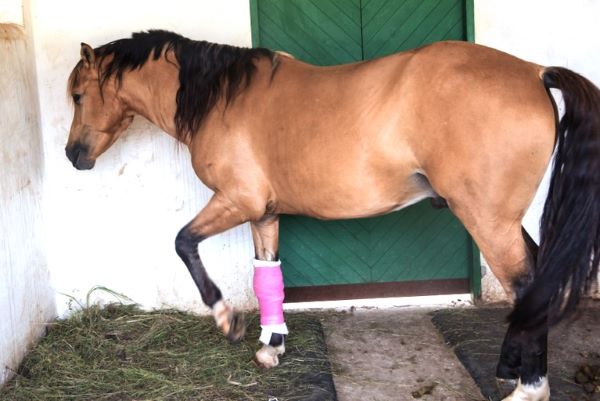
Even though sprains happen frequently, they may not always be immediately obvious. Often, there’s little noticeable issue during exercise, and it’s not until the evening when heat, pain, and swelling become evident. Therefore, checking the foreleg tendons at the back of the cannon bones typically forms part of the evening routine, especially for horses doing hard work.
Prompt rest is paramount in all cases of sprain in order to prevent further damage or internal bleeding within the tendon. The subsequent steps involve reducing the swelling through cold-hosing, application of ice packs, and physiotherapy equipment such as ultrasound or short-wave therapy. To provide support, it’s essential to bandage the legs, including the opposite leg likely to bear extra weight.
Advanced Treatments for Severe Sprains
With severe sprains, particularly of the foreleg tendons, a shoe with a raised heel may be fitted to provide support and relieve tension on the injured tendon. In some cases, a plaster cast may be needed to offer support and let the leg rest.
The final treatment stage centers around promoting healing. To encourage this, irritant lotions or pastes, known as “blisters,” are sometimes applied on the skin over the damaged tendons once the initial inflammation has subsided. These aim to increase the blood flow to the affected region, but their effectiveness remains a topic of debate. It’s theorized that the real benefit comes from the enforced rest following tendon treatment.
Various surgical procedures have been developed to improve and strengthen tendon healing. It’s crucial to consult your vet to determine if any of these interventions are advisable for a specific sprain.
Healing Time and Recovering from Sprains
Healing speed depends on the tissue’s blood supply. Muscles have an abundant blood vessel supply and heal quickly when injured. Tendons and ligaments, however, have a poorer blood supply, and healing in such tissues is extremely slow. A full recovery for a damaged tendon might take at least six months, perhaps longer. For ligaments, a recovery period of a minimum of nine months is expected. A spell at grass of 6-12 months is advisable for a tendon sprain, and at least 12 months for a serious ligament strain, such as a check ligament sprain.


